
Showcasing the complex and fascinating world of
medical research through captivating scientific images.
A Cellular Nebula
Dr Bobby Boumelhem
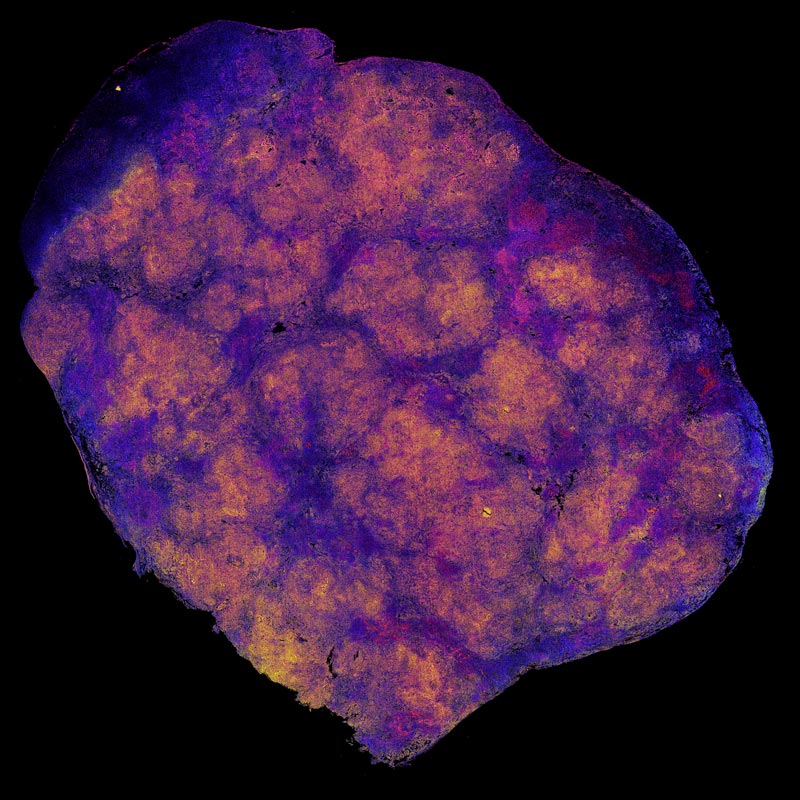
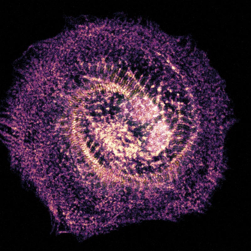
What resembles a beautiful, swirling cosmic storm is actually a detailed look at cancer’s chaotic growth. This vivid image, titled ‘A Cellular Nebula’, captures a microscopic view of a mouse liver affected by hepatocellular carcinoma, a primary form of liver cancer. The intense, fiery glow of pinks, purples, and oranges highlights dense clusters of rapidly dividing tumor cells. By transforming hidden biological details into vivid colour, the image helps researchers better understand how liver cancer develops and spreads — knowledge that may one day guide new treatments for patients.
Landscape of Renewal
Dr Mojdeh Abbasi
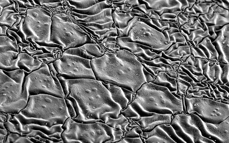
This scanning electron microscopy image shows corneal stem cells growing on a specially coated contact lens that supports limbal stem cells, which maintain and regenerate the cornea. Resembling a miniature landscape, the organised cells highlight a promising approach to repairing the ocular surface and restoring vision, inspiring the title Landscape of Renewal.
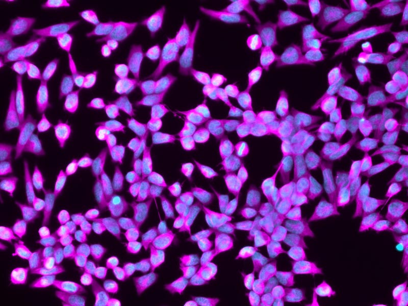
Vision in Bloom
Hannah Madanowski
This fluorescent microscopy image captures cultured corneal epithelial cells, the remarkable cells which play a crucial role in protecting the eye and maintaining the transparency needed for clear vision. These cells are being used to further our research into limbal stem cell deficiency, a condition in which the cornea’s ability to regenerate is disrupted, leading to blindness. Studying the characteristics of corneal epithelial cells helps us to gain new insights into the mechanisms of cornea and contribute to the development of regenerative therapies that can preserve vision.
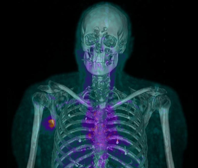
An Unprecedented Look at Tumour Biology
Professor Philip Hogg
Centenary researchers have developed a technology called CDI PET to image dying and dead cells in tumours for the first time. The image shows dying and dead cells in a skin cancer tumour in the right armpit of the patient.

Glacial Tears
Dr Felix Marsh-Wakefield
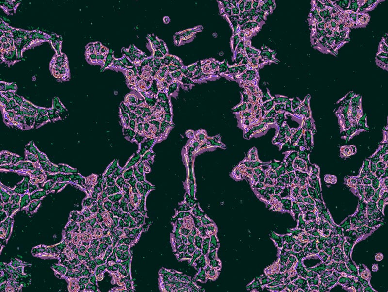
At first glance, this image appears to show a glacier carved by streams of meltwater. In reality, it is a liver tumour viewed through advanced imaging technology. The resemblance is more than visual. Both glaciers and liver cancer often change slowly over many years, yet each can reach a tipping point where progression accelerates and recovery becomes increasingly difficult. The silhouetted figures remind us that these changes are not merely scientific observations. They shape lives, futures, and the world we leave to the next generation.
Between Attachment and Escape
Dr Thuy Luong
Caught in a critical moment of transformation, these melanoma cells are preparing their escape. Though still anchored, they stretch outward to weave delicate connections. Soon, they will detach, gathering into resilient, floating clusters to travel through the body. Our research focuses on this exact shift—how cancer cells abandon their anchors to survive stress, migrate, and resist medication. By bringing this microscopic choreography to light, we decode the survival strategies of melanoma, bringing us one step closer to dismantling its defenses before it can spread.
The Bloom
Dr Angela Ferguson
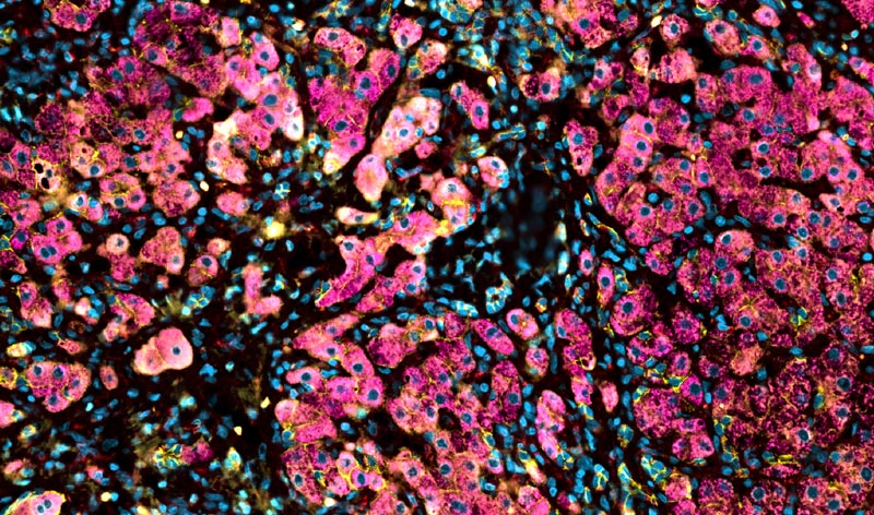
At first glance, The Bloom resembles vibrant pink blossoms, suspended in motion, as though drifting on water. Beneath this natural beauty lies a different reality. Cancer cells form striking pink structures contrasting dark surroundings, revealing a landscape shaped not by growth in harmony with nature, but by uncontrolled proliferation of disease. The Bloom embraces this tension between appearance and truth. Blooming, often associated with health and new beginnings, here, reflects a process of uncontrolled expansion, challenging view Bloom reconsider their assumptions about beauty. Transforming microscopic pathology into a landscape that speaks to the complexity and fragility of life and growth.
Into The Galaxy
Serena Li
Looking for the cause of inherited heart diseases in our patients is like exploring a vast galaxy of possibilities. Making our own heart cells and looking into the fine details of the cells is one way we start exploring. A single cell is the first step in understanding and answering the many questions about our patients’ diseases.
The Flutter of Breath
Guanshu Zhao
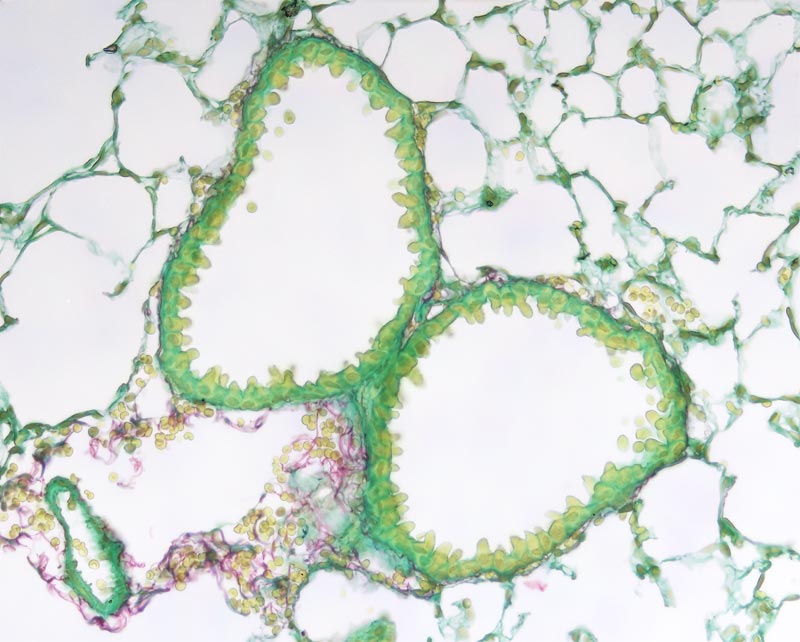
This image shows a delicate lung tissue section shaped like a butterfly, with two airways forming its symmetrical “wings.” The sample is stained using Sirius Red, highlighting the fine structural framework that supports breathing. Beyond its visual elegance, this structure reveals how intricately the lung is organised to sustain airflow and gas exchange. By combining biological form with an almost artistic symmetry, the image reflects the hidden architecture of respiration—where every breath depends on precisely arranged microscopic structures working in harmony to keep life in motion.
Constellations of a Dying Liver
Dr Bobby Boumelhem
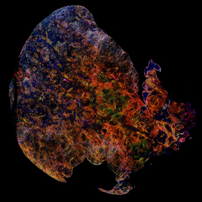
What looks like a cosmic storm is a close look at liver cancer in a mouse model, transformed through scientific imaging. This vivid microscopic landscape captures the hidden drama unfolding inside a diseased organ — a world shaped by growth, stress and survival. Fiery reds and glowing golds trace regions where the tumour and surrounding tissue interact, while cooler tones hint at subtle differences in structure. By visualising these changes, researchers gain new clues into how liver cancer forms and how it might one day be stopped.

The Snowfall before Liver Christmas
Dr Catherine Lamarque
The face of Jack from “The Nightmare Before Christmas” is shaped by the place where blood vessels and a bile duct come together in a transplanted donor liver. Around his face, the snow is made of cells normally present when the liver is healthy. Like Jack discovering a new world, we need to understand how the liver works in health before we can understand what changes when it becomes sick. By studying this precious donated liver, we hope to learn how to protect this fragile cellular snow and prevent it from disappearing.
Inflammatory Supernova
Hanna Gong
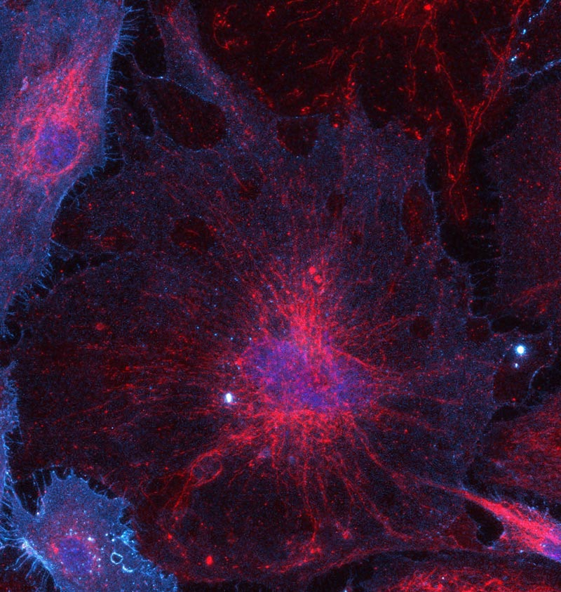
This image captures a senescent human umbilical vein endothelial cell (HUVEC) undergoing inflammatory and mitochondrial remodelling. The red mitochondrial network radiates outward like a stellar explosion, while blue and cyan signals highlight the nucleus and inflammatory activation surrounding the cell. Although visually reminiscent of a distant cosmic nebula, the image represents cellular ageing within blood vessels — a process linked to chronic inflammation and cardiovascular disease. By revealing the hidden beauty of microscopic biology, this work explores the striking visual connection between human cells and the universe.
Hold It Together
Serena Li
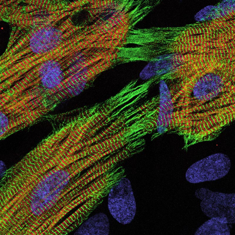
Heart muscle cells hold tight onto each other through three special links that couple the cells electrically and mechanically so that they beat in a controlled and coordinated manner. Even when there’s only a few of these cells, they work hard to maintain contact with each other so they can continue the communication required to work well together.
Mesenchymal Stem Cell Architecture
William Li
What appears at first glance to be a vibrant abstract artwork is in fact a community of mesenchymal stem cells viewed under a fluorescence microscope. These remarkable cells play an important role in tissue repair and regenerative medicine. By highlighting different structures within the cells using fluorescent markers, the image reveals a hidden landscape of intricate networks, glowing pathways, and striking colours. It offers a glimpse into the beauty of life at a microscopic scale, where scientific discovery and art naturally intersect.

Monday Mood
Dr Christine Lee
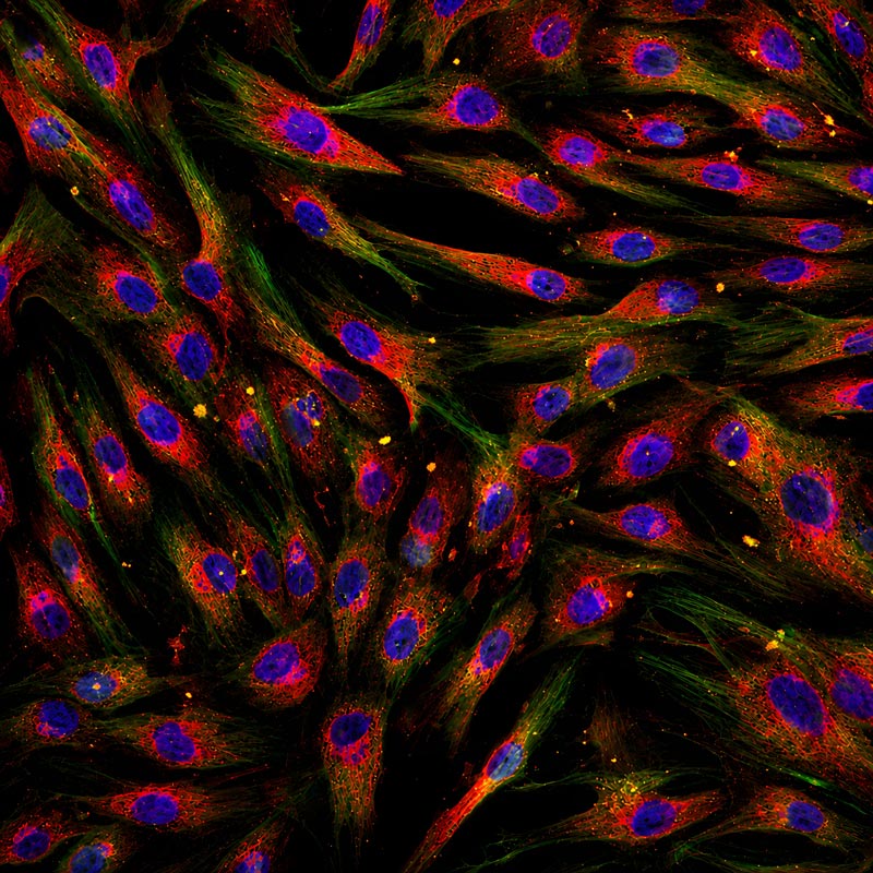
A little sleepy, slightly stretched, and just holding it together, this appendiceal organoid perfectly captures the feeling of a Monday morning. Each glowing blue dot is a cell nucleus, while the red outlines show the cells clinging into a neat, donut-like shape, as if forming a support group to get through the day. Grown in the lab to mimic real human tissue, this tiny structure helps scientists’ study how cells behave and respond to treatments, proving that even on Mondays, something productive is happening.
Blue Moon Rising
Dr Angela Ferguson
Blue Moon Rising through darkness, its surface marked by glowing craters, suggesting a new molten world arising. Hidden is a microscopic view of liver cancer. Tumour cells echo vibrant craters of intense biological activity, their warm hues contrasting against surrounding darkness. What appears serene and beautiful reveals a complex reality: the relentless growth and adaptation of cancer within living tissue. Blue Moon Rising reflects the paradox that disease can emerge from beauty. The image invites viewers to contemplate nature’s recurring patterns, hidden landscapes in the human body, and how art can transform disease into a moment of awe and reflection.

It’s Pawfect
Serena Li
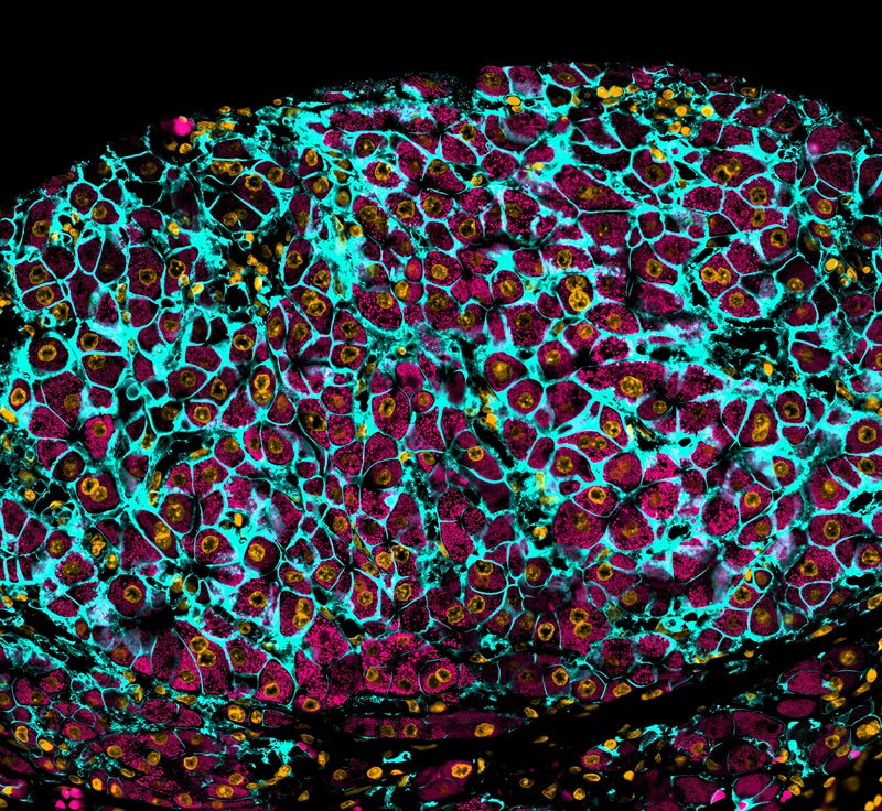
Cardiomyocytes are the beating cells of the heart, where every part highlighted in yellow and red work together to make them beat spontaneously. This is a ‘pawfect’ representation of the teamwork in our research group who work together to uncover the causes of sudden cardiac death that affect too many young people in our population.
The Opal of My Eye
Ka Ka Ting
This is an image of a mouse deep cervical (neck) lymph node which is covered in blood vessels in red and lymphatic vessels in green. Waste from the brain is cleared and filtered through these lymph nodes, where immune cells process it. These nodes act as biological “bins” and its dysfunction has been shown to contribute to Alzheimer’s disease. This research is focused on improving the clearance of waste by targeting blood and lymphatic vessels.

That’s No Moon
Dr Felix Marsh-Wakefield
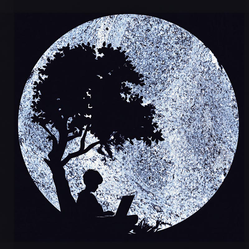
The moon is instantly recognisable no matter how it is photographed. For biomedical researchers, the tonsil is much the same. As one of the most studied and frequently used reference tissues, its distinctive structure is familiar across countless experiments and imaging techniques. In this artwork, a tonsil section becomes a moon illuminating a child beneath a tree, working on a laptop. It celebrates the curiosity that drives science: the ability to look at something familiar and see it differently. Even the most routine images can inspire new ideas, discoveries, and wonder.
Islands of Life in a Cellular Ocean
Dr Thuy Luong
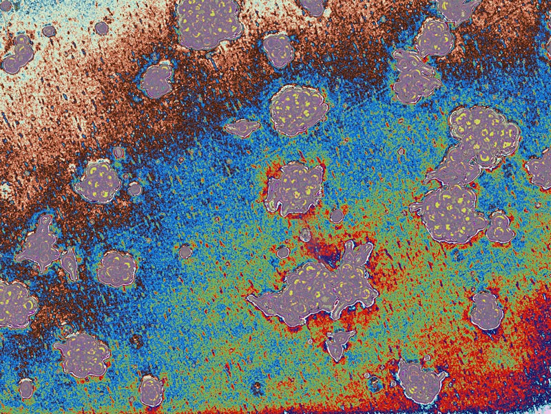
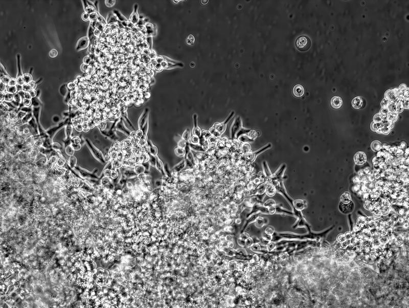
At first glance, this image looks like serene islands scattered across a quiet sea. In reality, it reveals a dangerous transformation. These are melanoma cells shifting their survival strategy. While some remain firmly anchored, others break free to form floating clusters. This shape-shifting ability allows the cancer to endure stress, travel through the body, and resist treatment. By tracking these microscopic changes, our research decodes how melanoma adapts and spreads. Understanding this critical transition is a vital step toward developing new, targeted therapies to stop the disease before it drifts to new shores.
Coral Reef
Dr Thuy Luong
This image captures melanoma, a dangerous form of skin cancer – in action. The vivid clusters you see are “spheroids,” tiny 3D spheres that cancer cells form to survive and prepare for their deadly journey through the body. This huddling process is a critical first step in metastasis, or how cancer spreads. By mapping exactly how these rogue cells group together and migrate, our research aims to decode the cancer’s playbook. Unlocking these secrets brings us closer to developing powerful new treatments designed to break these clusters apart and stop melanoma in its tracks.
Seeds Of Vision
Dr Mojdeh Abbasi
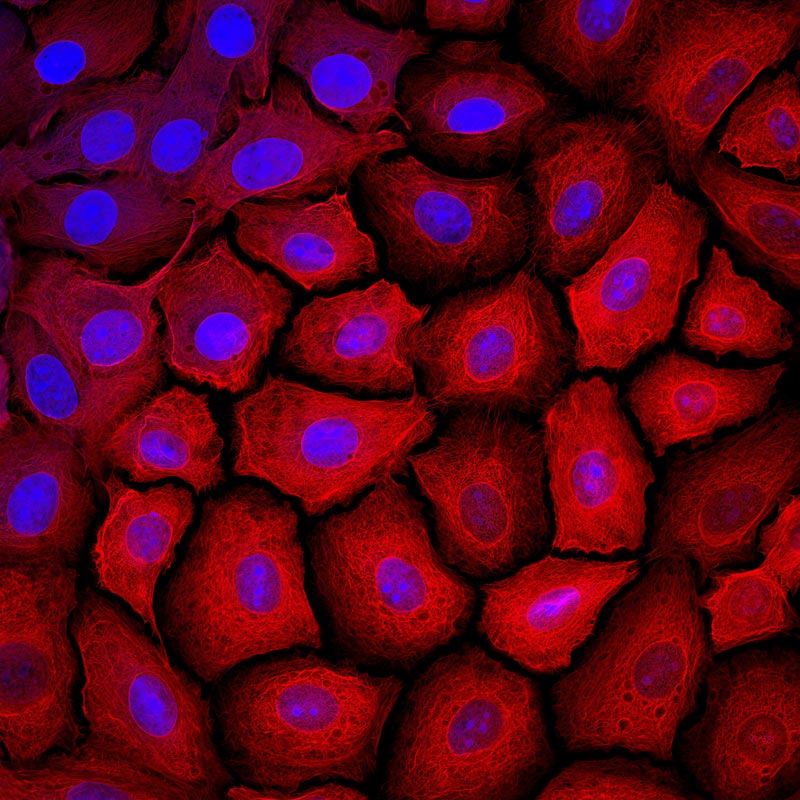
This image shows corneal stem cells cultured on a specially coated contact lens designed to support regenerative therapies for the eye. The cells are stained for Keratin 14, a marker associated with stem cells and visualised using fluorescence microscopy. These cells are being studied as part of a regenerative approach to repair the damaged surface of the eye. Inspired by their potential to restore sight.
A Bomb Hit My Desk Then a Dog Ate My Thesis
Jinx Moore
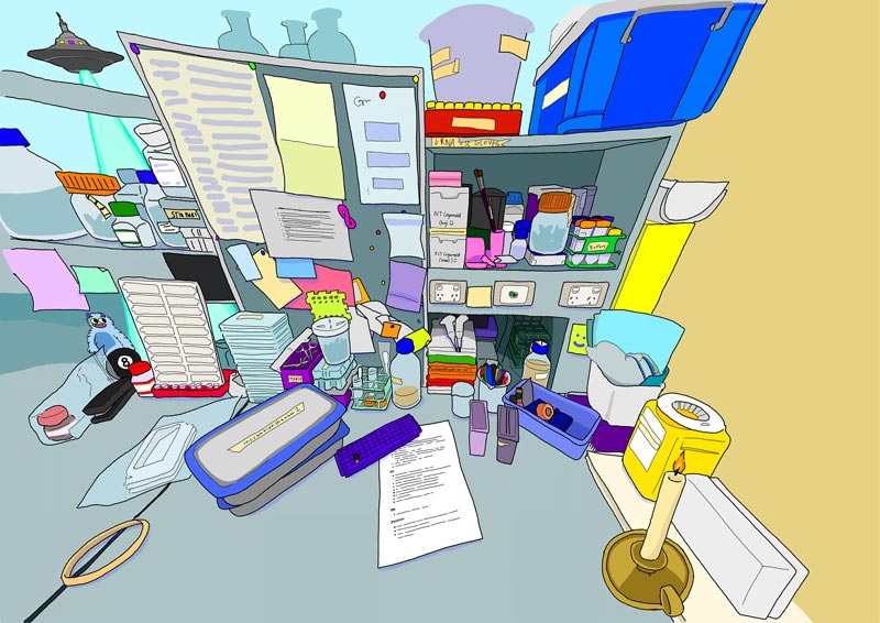
A bomb hit my desk then a dog ate my thesis (and other things that have definitely not happened) – Even though one’s bench may look crazy after an experiment, there is also a certain drama to the chaos. An everyday scene is presented in an unexpected way, depicting the bench where most experimental work takes place.
Peacockverse
Dr Chirag Parsania
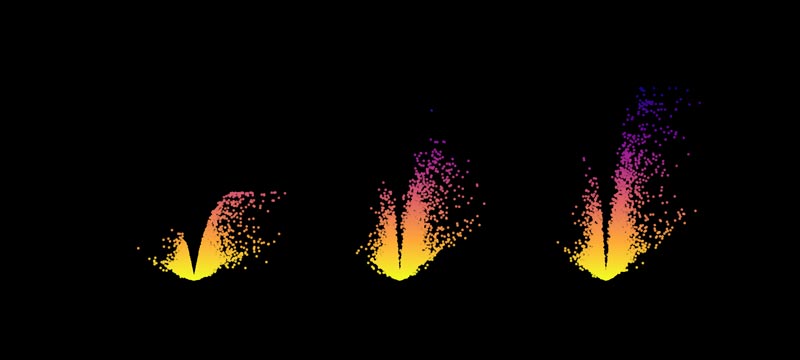
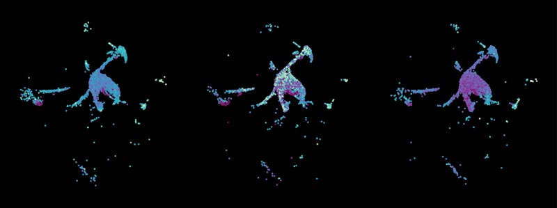
Peacockverse captures the hidden beauty of mouse lung cells in a study of a newly discovered gene involved in virus-triggered immune responses. Each point represents a single cell, arranged into peacock-like patterns across three genotypes: Wild-type, heterozygous and knockout. The bright colours highlight cells showing interferon-stimulated responses, a key defence mechanism against viral infection. By transforming complex single-cell data into an accessible visual story, this image reveals how genetic changes can reshape immune activity in the lung.

Fault Lines
Dr Bobby Boumelhem
This image reveals the hidden architecture of a diseased liver in a mouse model of liver cancer. Fine strands of tissue glow like illuminated fault lines, tracing areas where scarring has reshaped the organ. These structural changes, often invisible to the naked eye, can influence how tumours grow and how the liver functions. While these complex patterns look like a beautiful, glowing constellation in the night sky, they map the biological struggle against disease. By studying these microscopic environments, researchers gain vital insights into how tumours grow, bringing us closer to discovering more effective treatments.
Volcano of Viral Defence
Dr Chirag Parsania
Volcano of Viral Defence transforms gene activity in the mouse lung into a striking volcanic landscape. Each dot represents a gene, shown across different genetic backgrounds of a newly discovered gene involved in antiviral immune defence. The eruption-like pattern highlights genes linked to interferon responses — one of the body’s key early warning systems against viral infection. By turning complex genomic data into a vivid visual story, this image reveals how genetic changes can reshape the lung’s immune response to viruses.