
Showcasing the complex and fascinating world of
medical research through captivating scientific images.
Thermal Tapestry –
Dr Bobby Boumelhem
(People’s Choice winner)
This vibrant image reveals the hidden beauty of brown fat, a special type of fat that burns energy to keep us warm. Taken from a mouse and captured using confocal microscopy, the tissue was stained to highlight its microscopic features: lipid droplets glow magenta, free fatty acids shimmer in blue and cell nuclei shine yellow. Unlike white fat, brown fat is packed with mitochondria and fuels heat production—a natural internal heater. This glimpse into the cellular landscape helps scientists understand how brown fat works and how it might one day be harnessed to combat obesity and metabolic disease.

Dark Transit –
Dr Angela Ferguson
(1st Prize – Judges’ Choice)
From above, the image evokes a familiar calm: glowing threads of light slicing through darkness, a highway at night—orderly, rhythmic, alive. But this is not city infrastructure. These illuminated paths are not roads, but vessels hijacked. Not cars, uncontrolled cells traveling routes they should never take. The network, once purposeful, now corrupted for silent invasion. Dark Transit illustrates how liver cancer spreads: silently, systematically, beneath the surface, weaving changes in the body’s internal landscape flowing through an organ unaware it’s being overtaken. What appears efficient and structured becomes a map of disruption—an anatomy of invasion cloaked in familiarity..
False Bloom –
Dr Angela Ferguson
At first glance, a radiant field of sunflowers stretches endlessly—each bloom golden, full of life, stretching skyward. But look closer. The symmetry is too perfect, the growth too aggressive. These flowers do not follow the sun—they devour it. Petals fray into twisted strands, crowding over one another in a dense, consuming sprawl. This is not a celebration of life, but a camouflage for something darker: a landscape of unchecked growth, of beauty weaponized. False Bloom is an image of liver cancer—how it can mimic vitality even as it undermines it, turning invasive and altering something once beautiful.


Heartstrings in Fluorescence –
Dr Bobby Boumelhem
This glowing image offers a rare glimpse inside a mouse heart captured using confocal microscopy. Stained to reveal its intricate structure, magenta highlights lipid content, blue indicates other cellular components and white pinpoints the nuclei—the command centres of each cell. Far from just a pump, the heart is a dynamic, energy-hungry organ. By visualising its microscopic landscape, scientists can uncover how fats are stored and used in cardiac cells—insights that may help us understand heart health and disease at the most fundamental level. It’s science at the crossroads of structure, function and beauty.
Yummy Spread –
Nguyen Dac Thuy Luong
Melanoma is one of the most aggressive skin cancers, often spreading quickly throughout the body. My research focuses on a specific melanoma cell line, YUMM3.3, to understand how these cancer cells form 3D tumour-like structures (spheroids), detach from their original site and migrate to distant organs. By studying these behaviours, we aim to uncover the key mechanisms that drive melanoma’s deadly ability to metastasise. This knowledge could pave the way for developing more effective treatments that prevent cancer spread and improve patient survival. Here we see the YUMM3.3 cell line forming spheroids, detaching from the primary site exhibiting metastatic behaviour..


Confetti Snowdome –
Dr Mojdeh Abbasi
(2nd Prize – Judges’ Choice)
This image shows the mouse cornea from a Confetti reporter line after Cre recombination, with specific cornea stem cells labelled in distinct fluorescent colours. The mosaic pattern allows us to trace clonal expansion and track limbal stem cells. This approach reveals how stem cells contribute to corneal maintenance and regeneration over time. The vibrant, suspended colours evoke the appearance of a snow dome, capturing a frozen moment of cellular movement and regeneration.
Unseen Currents:
Sudden Death in the Young –
Yuchen Chang
(3rd Place, Judges’ Choice)
This artwork uses the R package “aRtsy” to visualise the silent and unpredictable nature of sudden cardiac death in young individuals. Inspired by Tyler Hobbs’ Flow Fields algorithm, the image represents a stream of individual life trajectories, each influenced by subtle, often undetected forces—genetic, environmental or unexplained. Each dot represents a sudden death patient with the angle field guiding the flow based on age data. These invisible currents symbolise how lives are shaped, disrupted or cut short by unseen factors. As lines meander, twist and converge, the piece evokes life’s fragility and complexity, where direction can change without warning.

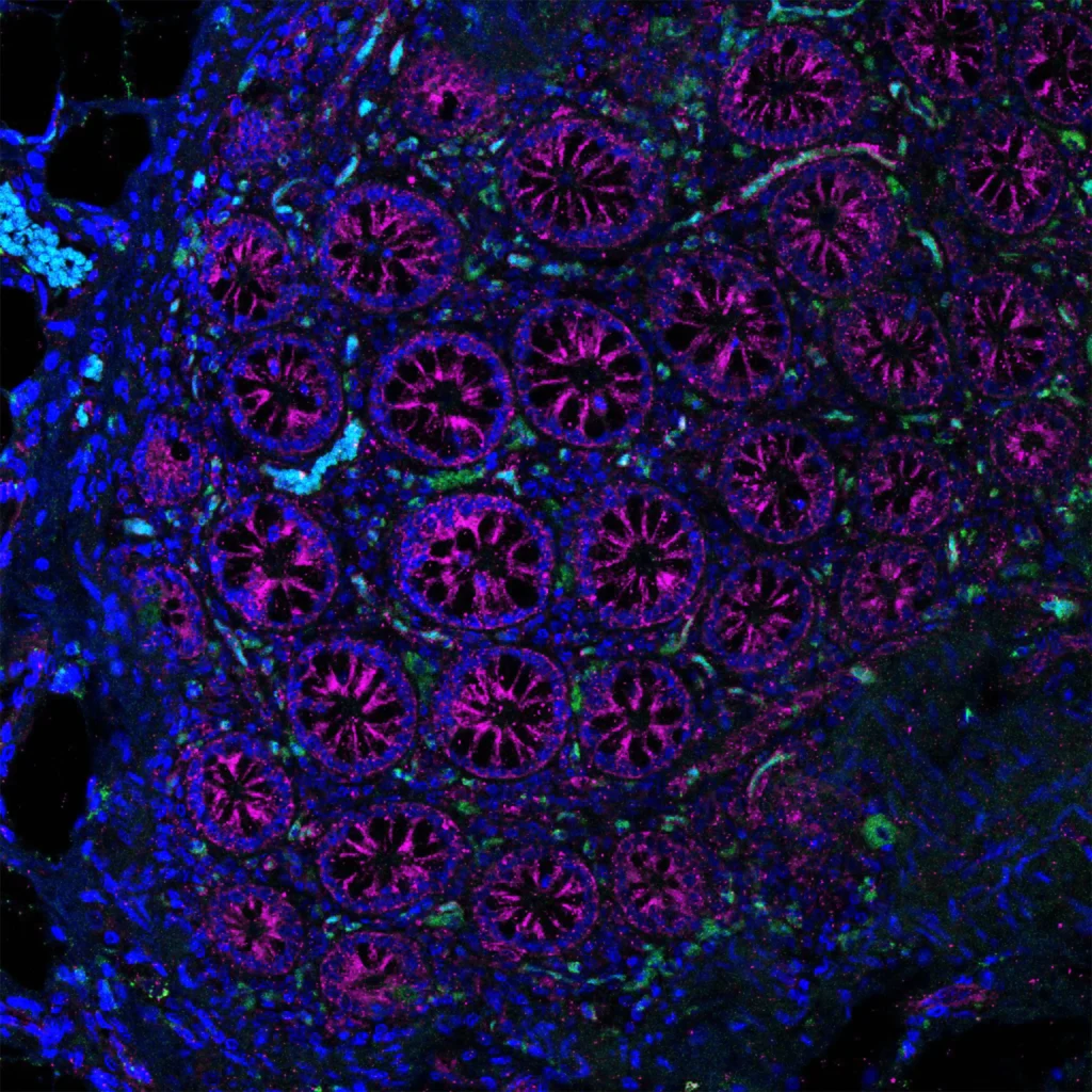
Colon Kaleidoscope –
Dr Bobby Boumelhem
(Staff Pick winner)
This striking image unveils the inner workings of the colon in a diabetic non-human primate, captured through the lens of confocal microscopy. Stained for key cellular players, magenta marks fibroblast activation protein (signalling tissue remodelling), while green highlights CD68, a marker of immune cells responding to inflammation. Blue shows the nuclei, the control centres of each cell. Together, these glowing signals paint a vivid picture of how diabetes may alter gut health. By studying these cellular changes, researchers aim to better understand the gut’s role in chronic disease and how healing might begin at the microscopic level.
The Flame In Between –
Natnicha Ketchaikosol
Amid a field of transformation, two cell types — epithelial and mesenchymal — reveal their distinct identities through fluorescent staining. Sourced from head and neck cancer, the mesenchymal cells, marked by vimentin, glow like a flame in a forest, quietly blazing through the structured canopy of epithelial order. This image captures more than cellular difference; it reveals the space where change ignites. This image reflects transition, conflict and the quiet fire within — a visual metaphor for the unseen forces driving transformation in life and disease.


Threads of Sight –
Dr Mojdeh Abbasi
This image, captured using scanning electron microscopy (SEM), reveals the finely textured surface of a specially coated contact lens, patterned like a woven fabric. Far more than a visual motif, we hope that this intricate design supports the growth of limbal stem cells—vital cells that naturally regenerate the corneal surface. When these cells are lost due to injury or disease, the result can be painful vision loss or blindness. Our research explores a patient-specific therapy that grows these cells directly on contact lenses. Once placed on the eye, this treatment offers the potential to restore sight and revolutionise treatment.
Pathways to a Broken Heart –
Yuchen Chang
This maze, shaped like a broken heart, was generated entirely in the R programming language using math functions and computer-aided machine learning. When art meets science, solving the maze becomes a metaphor for the complexity of researching genetic heart disease. Navigating its twists and turns can be both confusing and encouraging, much like the journey through genomic heart disease research. This work invites us all to think: can we code our way through the challenges of genetic disease?


Cellestial Nebula –
Natnicha Ketchaikosol
This image captures a 3D spheroid composed of epithelial and mesenchymal cells derived from head and neck cancer. Acquired through Z-stack imaging and rendered as a maximum intensity projection, the structure resembles a nebula in deep space—glowing, layered and full of dynamic complexity. Fluorescent staining reveals cellular diversity and organisation, reflecting the transitional nature of cancer and the silent forces shaping its evolution.