2024 When Art Meets Science
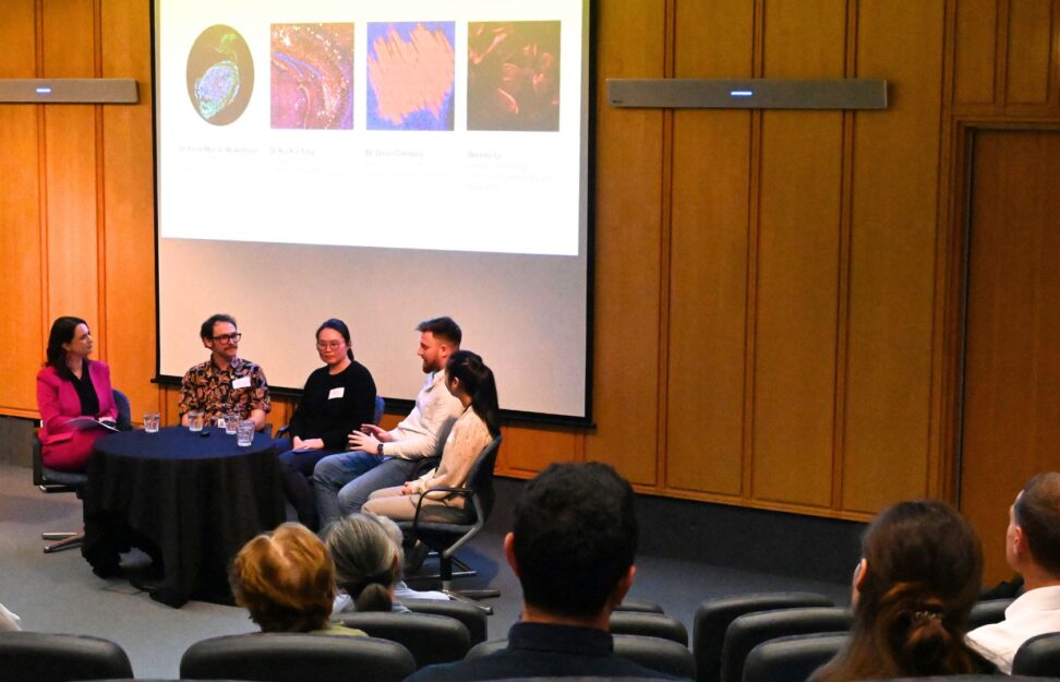
Centenary is delighted to have welcomed members of the community to the Institute for ‘When Art Meets Science’ as we celebrated National Science Week. Showcasing the complex and fascinating world of medical research through captivating scientific images our scientists shared the inspiration and science behind their artistic masterpieces.
Judged Category Awards
Determined by a panel of external judges we congratulated the winners of the annual Image Prize for their striking images.
-
First Place
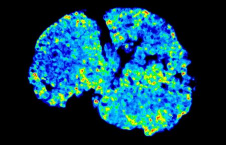
Cloudy Brain with a Chance of Forgetfulness – by Ka Ka Ting
-
Second Place
Close To My Heart – by Bobby Boumelhem -
Third Place
Roses are Red – by Heidi Strauss
This year’s entries
-
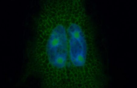
A Bridge Between Worlds – by Bobby Boumelhem
Pluripotent stem cells hold immense promise for regenerative medicine. They can be used to make any cell in the body. Here, we made neurons—the functional cell of the brain—from mouse pluripotent stem cells. By understanding how they develop, we can bridge the gap and mimic these processes in human pluripotent stem cells with the aim of generating or possibly replacing damaged or diseased neurons. -
Bed of Roses – by Heidi Strauss
Pancreatic cancer is no bed of roses, yet here it is. We are investigating pancreatic cancer utilising cells grown directly from donated patient tumour tissue and hope to identify novel therapies that target this devastating disease. -
Celestial Luminescence – by Hanna Gong
‘Celestial Luminescence’ captures the intricate beauty of cellular structures, where the vivid colours represent various components within a single cell. This image shows our research into mitochondrial structure, function and cellular health. By visualising these microscopic elements, we unlock new understanding of how cells operate, potentially leading to breakthroughs in treating age-related diseases and enhancing overall human health. The fusion of art and science in this image not only highlights the complexity of life at a cellular level but also invites viewers to appreciate the elegance of biological processes. -
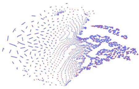
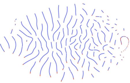
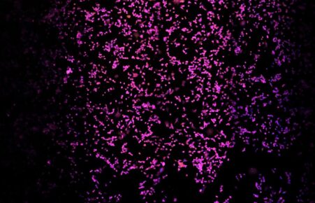
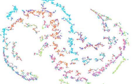
Cellular Cosmos – by Yew Wong
This artistic visualisation captures the intricate beauty of cellular structures. The blue and red dots, derived from analysing the characteristics of cellular images, create a dynamic pattern that suggests both complexity and movement. The arrangement forms a three-dimensional illusion, with a swirling motion that draws the eye towards a central point on the right. The dense clusters of dots indicate areas of intensity, while the sparser regions evoke a sense of expansion, reminiscent of a vortex or galaxy. This piece beautifully merges scientific analysis with artistic expression, highlighting the elegance of cellular organisation. -
Cellular-Honeycomb – by Keshav Raj Paudel
Imagine a cell’s interior as a dazzling honeycomb, where nature’s architectural marvel meets microscopic precision. Each hexagonal cell represents a vital unit of life, meticulously designed to support cellular functions and structure. This image unveils the intricate and elegant framework that keeps our cells functioning smoothly, showcasing the beauty and complexity of life on a scale that’s both fascinating and essential. Through this lens, we glimpse the seamless blend of science and artistry that underpins our biological world. -
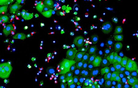
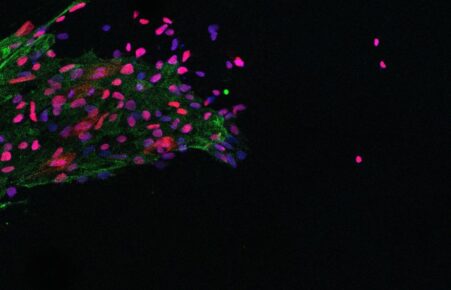
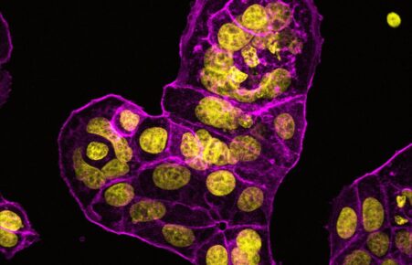
Close to my Heart – by Bobby Boumelhem
Approximately 60 women are diagnosed with breast cancer every day. Despite early screening and intervention providing good outcomes, much remains unknown about the machinations driving breast cancer. In this image, cells taken from a breast cancer tumour were grown in a dish to visualise how these cells change under different conditions. If we can piece together what occurs at the molecular level, we may gain much needed insight into this horrible malignancy. -
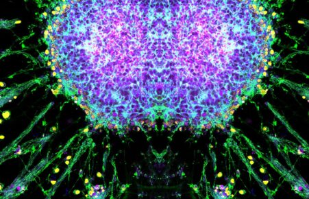
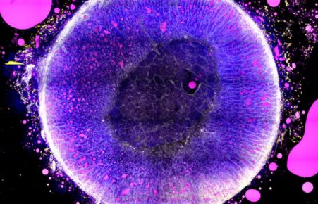
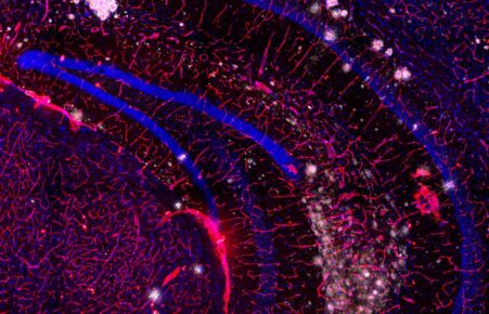
Cloudy Brain with a Chance of Forgetfulness – by Ka Ka Ting
This image shows the region of hippocampus in the mouse brain, which is riddled with round and fluffy-looking amyloid plaques (white). These plaques can damage the blood vessels (red), which provide a selective barrier to filter out harmful substances in and out of the brain. The hippocampus is an important storage hub for our memories, thus amyloid damage to both the brain cells (blue) and blood vessels can lead to loss of memory, a devastating symptom often seen in Alzheimer’s disease patients. -
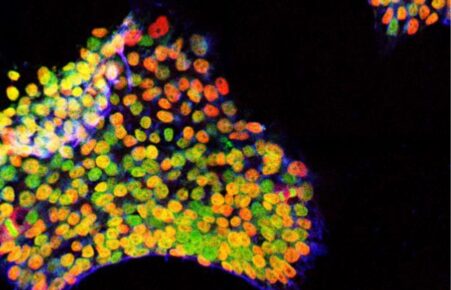
Colorful iPSC in the shape of Australia – by Anyapat Atipimonpat
This image shows a colony of induced pluripotent stem cells (iPSCs) arranged to mimic the shape of the Australian continent. The colony’s vivid colours represent different expressions of pluripotent stem cell markers, reflecting the dynamic nature of these cells. This colourful display not only symbolises the diverse and vibrant populations that inhabit Australia but also highlights how these tiny cells contribute to scientific advancements and medical breakthroughs around the world. -
Dodgem CARs – by Heidi Strauss
This image shows pancreatic cancer cells (green) being targeted and destroyed by modified immune cells known as CAR-T cells (red). We are currently investigating several different targets that are specific to pancreatic cancer with the hope of providing an effective treatment option for patients with this devastating disease. -
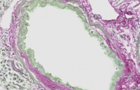
Gazing Doughnut – by A’qilah Banu
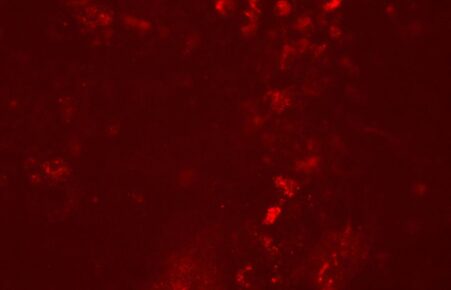
The gazing doughnut showcases the lung airway of a mouse, which has increased collagen deposition, depicted in red. This increased deposition causes scarring of the lungs which impedes the flexibility of the airways. This causes difficulty in breathing, frequent coughing and discomfort. This shows that the airway is not in optimal condition for efficient gas exchange, preventing the lung from functioning at its maximum capacity. Increased red deposition also causes airway stiffness and remodelling of the airway. Collagen synthesis is pivotal in dictating tissue regeneration, maintaining the structural integrity of healed tissues and matrix remodelling. -
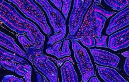
Grand Canyon – by Jasmine Minh Hang Nguyen
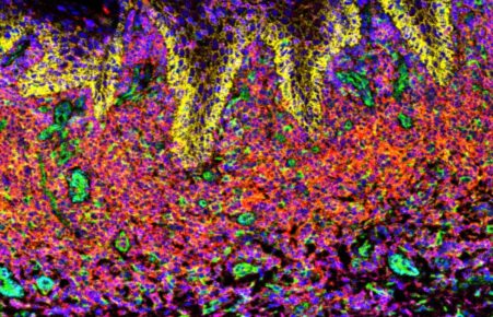
This artwork captures the cross-sectional view of the intestine from a mouse. With its vibrant colours and intricate patterns, the image mirrors an iconic landscape from an aerial view – the Grand Canyon. Each intestinal crypt (purple) is packed with cells (pink) and neatly organised side by side, resembling the mysterious and maze-like terrains waiting to be explored. Through this piece, I hope the viewers can appreciate the mesmerising depths and contours of Mother Nature, at a cellular level. -
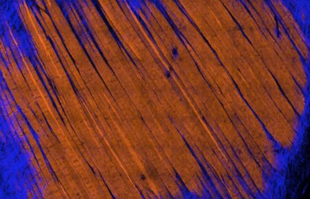
Great Sunburnt Land – by Dean Campelj
Here we demonstrate how muscle fibres and mitochondria, the energy cells of our body, are organised in an intertwined lattice structure (orange/brown), where the muscle fibres reside under connective tissue that holds them together in bundles (blue). The muscle fibres appear in the shape of Australia with the ocean surrounding, hence the name inspiration ‘Great Sunburnt Land’. This image is a still collage of a time-lapse video, acquired through intravital microscopy. This approach allows us to live image muscle fibres and mitochondria in real time providing the unique perspective we present. -
Heart of the Swarm – by Joseph Vitale
Overexpression of mutant adeno-associated virus (AAV) receptors in cancer cells. We are trying to understand how these receptors are localized and expressed in cells in the hopes of better understanding AAV transduction. Uncovering the mechanisms behind AAV transduction will allow us to improve the efficiency and outcome of clinical gene therapies in patients with genetic diseases. -
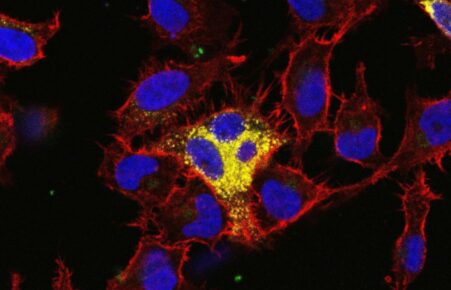
In the spotlight – by Althea Bastian
Pictured is a single cancer cell, identified from amongst millions of white blood cells, which is in the process of being picked by a needle slightly larger than the cell itself. By ‘wiggling’ the needle the cancer cell is dislodged and the surrounding cells are cleared away, allowing the cell to be taken up and further analysed. This delicate procedure allows us to investigate cancer cell biology on a single-cell level, and correlate that data to patient outcomes. -
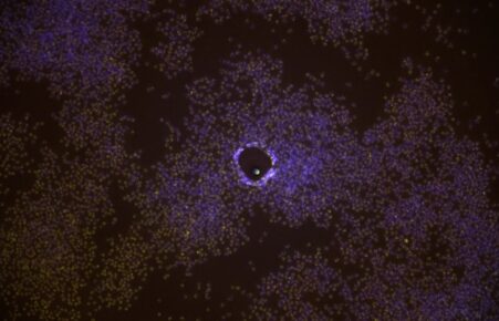
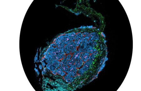
Lunar Lacus – by Angela Ferguson
Looking at healthy and diseased human tissue down a microscope is like looking at a map or in this case a lunar landscape. The large lakes of tumour are surrounded by cell-rich skin tissue. Immune cells have failed to infiltrate the tumour which has employed its defences allowing it to create ‘lakes’ of protected niches in the skin landscape. -
The Night Sky – by Nguyen Dac Thuy Luong
Head and neck squamous cell carcinoma (HNSCC) is a challenging cancer often driven by genetic mutations. Among these, HRAS mutations play a pivotal role. By transfecting cells with an HRAS plasmid, I delve into the genetic orchestration of this aggressive disease. HRAS, a proto-oncogene, regulates cell division and differentiation. When mutated, it can lead to uncontrolled cell proliferation, fuelling cancer growth. My research aims to unravel the precise molecular mechanisms of HRAS mutations in HNSCC, potentially uncovering new therapeutic targets. By understanding how HRAS drives cancer progression, we edge closer to innovative treatments and improved patient outcomes. -
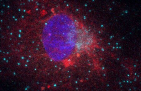
Molecular Constellations – by Hanna Gong
This image, resembling a cosmic night sky, actually depicts cells viewed through a fluorescence microscope. The colours highlight various cellular structures: the purple areas are cell nuclei, while red and orange networks signify mitochondria. Blue dots mark surface adhesion molecules E-selectin. This ‘molecular constellation’ offers a glimpse into the intricate beauty of cellular life, blending art and science to reveal the hidden wonders of our biological world. Just as a horoscope in the cosmos can guide you, this ‘molecular constellation’ can provide guidance in the diagnosis and treatment of certain diseases. -
Mountain Flames – by Angela Ferguson
To the human eye inflamed skin appears red and damaged, microscopically, inflamed skin appears as mountains of flames. The structural building blocks of our most important barrier, the skin, have been damaged by a fire-storm of immune cells tearing through its foundations. The question is, which cell started the fire and why? -
Neoplastic Nebula – by Natnicha Ketchaikosol
This image is a vivid 3D representation of a head and neck cancer cell line, showcasing the intricate interplay between epithelial and mesenchymal cells. It highlights cellular diversity and structure, offering a mesmerising glimpse into the microscopic world of cancerous growth. Remarkably, the image’s resemblance to the human brain underscores the complex beauty and unexpected patterns found within malignant transformation. This fusion of art and science encapsulates the enigmatic parallels between pathological and anatomical forms. -
Neural Symphony – Unveiling Cellular Secrets – by Yew Wong
Imagine peering into a vibrant, microscopic universe where cells dance in intricate patterns, mirroring the complexity of the human brain. By analysing images of cells, we unveil hidden behaviours and group them into fascinating clusters. This visual symphony, resembling neural networks, not only captivates the eye but also unlocks secrets of cellular function and organisation, paving the way for groundbreaking discoveries in biology. -
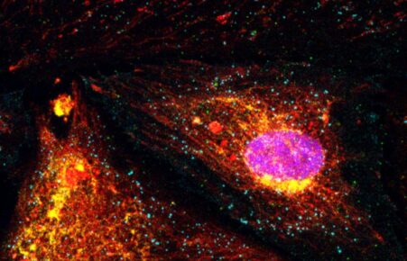
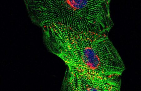
Peas in a Pod – by Serena Li
To study heart disease in a dish, these heart muscle cells were made from patient blood samples. Like peas in a pod, heart muscle cells like to stick together. They form strong bonds with their neighbours through a series of proteins, one of which is called connexin (shown in red). Connexins are responsible for transferring electrical signals between the cells and may be compromised in diseases like hypertrophic cardiomyopathy. -
Release – by Serena Li
This image shows stem cells derived from patient blood samples. These cells have the potential to turn into any cell type and have been instrumental in furthering patient-specific research and therapy development. By generating heart cells from these stem cells, we can study genetic heart diseases more effectively, getting us closer to releasing our patients from the unknown and uncertainty surrounding underlying disease mechanisms. -
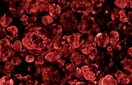
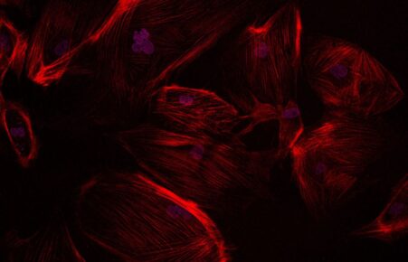
Roses are Red – by Heidi Strauss
“Roses are red, Cancer is blue, With perseverance, We’ll find a way through… In this image we are investigating pancreatic cancer utilising 3D cells grown directly from donated patient tumour tissue. Growing these cells in 3D provides us with patient-specific models that retain their complex architecture, functions and genetic profiles and are also long-term solutions to reducing animal research. This image shows the structure of these cells, which resembles a delicate rose. -
Roses from the Heart – by Serena Li
This image shows heart muscle cells generated from patient samples. The striations of the muscle cells resemble delicate and beautiful rose petals that fall from the flower after they bloom. By studying these cells, we can uncover the workings of genetic heart diseases to give hope and answers to our patients. -
Seedling of Truth – by Felix Marsh-Wakefield
Eighty percent of Australians diagnosed with liver cancer will die within five years. For Indigenous Australians, the mortality rate is even higher at 90%. These harrowing statistics starkly contrast with the serene image of the tumour resection, resembling a delicate seedling. This image underscores the intricate structure of liver cancers, showcasing their beauty amidst the brutality. Cancer is daunting, but by facing our fears we can improve our ability to study and conquer it. The seedling is blooming with secrets to be uncovered to improve survivability and transform liver cancer from a death sentence into nothing more than a pretty picture. -
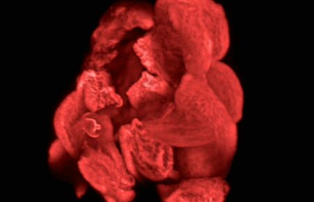
Somewhere in Another Universe… – by Long Chung
In another universe inside an mouse embryo, these bright stars driven by a developmental gene are travelling through millions of years to differentiate themselves into specified neuronal cells to regulate the movement of a mature mouse. -
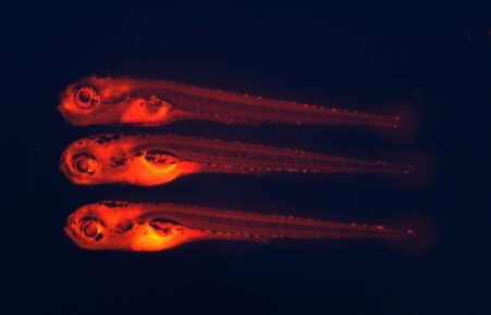
The Glowing Fishes – by Christelle Tshamala
The glow that you see radiating from the zebrafish in this fluorescence image will provide a baseline on how we can treat the early reversible stages of fatty liver disease, before it progresses into a chronic state. The fish have been treated with Nile Red (a fat soluble dye) attached to nanoparticles to help track differing fat levels across treatments. -
Tiny but Mighty – by Bobby Boumelhem
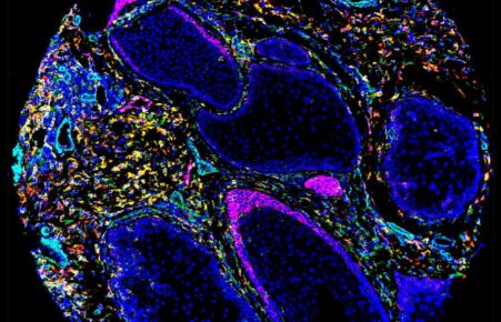
The adrenal gland is a small gland situated above the kidney. Despite its small size, this gland is responsible for releasing hormones throughout the body. Each section of the gland is responsible for releasing different types of hormones – from glucocorticoids that are pivotal in metabolism, to the stress hormone cortisol and sex steroid hormones like testosterone and estrogen. In this image, the adrenal gland was stained with lipid (fat) probes to better understand how lipids can affect its function. -
Yin and Yang – by Yew Wong
In this innovative study, we analysed cellular images, processing up to 256 unique characteristics to categorize cells based on their behaviours. The resulting visualisation, reminiscent of the Yin Yang symbol, beautifully illustrates the balance and harmony within cellular interactions. This approach not only enhances our understanding of cellular functions but also presents complex data in an accessible and visually captivating manner, bridging the gap between science and art. -
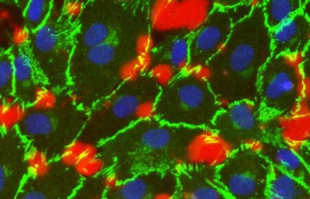
You Shall Not Pass! – by Ngan Ching Cheng
The endothelium is the inner lining of blood vessels. It is a layer of tightly organised endothelial cells. It forms a barrier to control the passage of fluid and molecules between the vessels and the surrounding tissues and so maintains vascular integrity. When the endothelium is compromised, it can trigger critical pathological conditions. In this image, endothelial cells in culture are shown. The red areas depict leakage among cells while the cell junctions are shown in green. When the junctions are intact, they form an impermeable barrier. When the junctions are interrupted, the leakage can be detected.